
MACULAR DEGENERATION
Age-related macular degeneration (AMD) is one of the leading causes of irreversible vision impairment in Australians over 55 years (1-3). The condition affects the macula, a specialised region at the centre of the retina responsible for the detailed central vision required for reading, driving, recognising faces, and performing many everyday tasks (4). Whilst peripheral vision typically remains unaffected, damage to the macula can profoundly impact independence, mobility, engagement in social activities, and mental aptitude and quality of life (5,6). Over recent decades, treatment advances have prolonged central clarity and dramatically improved visual outcomes for those with neovascular (wet) AMD, and contemporary treatments for geographic (dry) AMD show promise (7,8).
Whilst the immense impact of macular degeneration cannot be underestimated, thorough explanation of the condition, compassionate professional management, and optimisation of remaining vision have evidenced positive impacts on quality of life. Nicola has prior experience working alongside Vision Australia, and combines her expertise in diagnosing and managing pathology with her empathetic explanation of low vision correction options. By arranging a low vision eye examination at Onèj Optometry, you can ensure that not only will your vision and ocular health be thoroughly assessed with advanced retinal scans, but your options for treatment and management will be explored in detail.
-
AMD is a complex disease influenced by a combination of ageing, genetic predisposition and environmental factors. Smoking is the strongest modifiable risk factor, substantially increasing both the likelihood of developing macular degeneration and the rate at which it progresses (9-11). Other risk factors include increasing age, family history, cardiovascular disease, hypertension and prolonged exposure to ultraviolet radiation (12-14). Although many patients assume that vision loss is an inevitable consequence of ageing, early identification of disease and appropriate management can significantly reduce the risk of severe visual impairment. As such, regular, thorough eye tests are imperative.
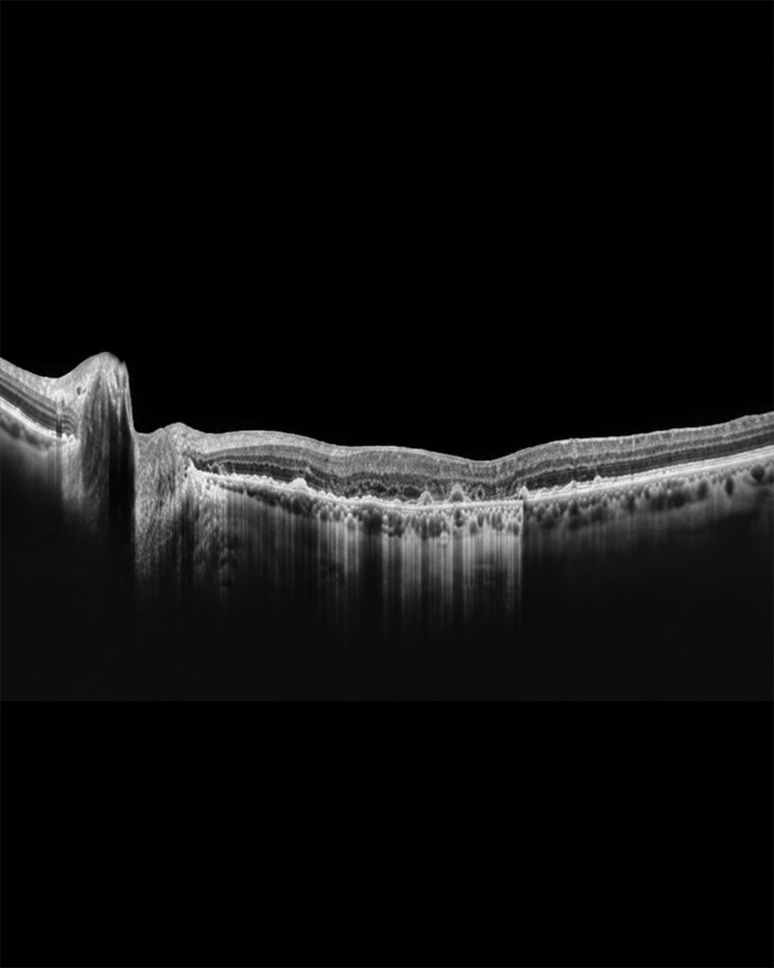
The earliest stages of macular degeneration are characterised by the accumulation of deposits known as drusen beneath the retina. These deposits are often accompanied by dysfunction of the retinal pigment epithelium, a layer of cells responsible for maintaining the health and metabolism of the light-sensing retinal tissue . In many patients, these changes progress gradually over many years and may initially cause few or no symptoms. As the disease advances, however, patients may experience distortion of straight lines, reduced contrast sensitivity, difficulty reading, impaired recognition of faces, reduced performance in dim lighting conditions, and blurred or missing areas within their central vision.
Macular degeneration is broadly classified into two forms. Dry AMD is the more common subtype and is characterised by progressive degeneration of retinal tissue at the macula. Although progression is often slow, advanced forms of dry AMD can result in significant and permanent central vision loss. Wet AMD is less common but typically more aggressive. In this form of the disease, abnormal blood vessels develop beneath the retina, resulting in leakage of fluid, blood and inflammatory mediators into the macular tissue. Without prompt treatment, these changes can cause rapid and irreversible deterioration of central vision(16, 17).
Because many of the earliest retinal changes occur before symptoms become noticeable, regular eye examinations are critical for early detection. Comprehensive assessment of macular health involves detailed retinal examination, retinal photography and optical coherence tomography (OCT), a highly sophisticated imaging technology capable of detecting microscopic changes within the retinal layers. These investigations allow subtle disease progression to be identified long before substantial vision loss occurs, facilitating timely intervention and ongoing monitoring.
At Onèj Optometry, we understand that a diagnosis of macular degeneration can be confronting. Our approach focuses upon thorough assessment, clear communication and evidence-based management tailored to the individual. We carefully evaluate each patient's risk factors, retinal findings and visual requirements before discussing appropriate monitoring intervals, lifestyle modifications and treatment recommendations. For patients with signs of disease progression, we collaborate closely with local ophthalmologists and other healthcare practitioners to ensure timely referral and coordinated care.
Preserving vision requires a proactive approach. Smoking cessation, cardiovascular health optimisation, appropriate dietary intake, ultraviolet protection and regular ocular examinations all play important roles in reducing the risk of vision loss associated with macular degeneration(17-19). Through comprehensive retinal assessment and personalised management, early detection of disease progression is possible, providing the greatest opportunity to preserve visual function and maintain quality of life for years to come.
References
1. Ly, A., Herse, S., Williams, M. A., & Stapleton, F. (2025). Artificial intelligence for age‐related macular degeneration diagnosis in Australia: A Novel Qualitative Interview Study. Ophthalmic and Physiological Optics, 45(6), 1282-1292.
Link: https://onlinelibrary.wiley.com/doi/full/10.1111/opo.13542
2. Guymer, R. H., & Campbell, T. G. (2023). Age-related macular degeneration. The Lancet, 401(10386), 1459-1472.
Link: https://www.thelancet.com/journals/lancet/article/PIIS0140-6736(22)02609-5/abstract
3. Keel, S., Xie, J., Foreman, J., Van Wijngaarden, P., Taylor, H. R., & Dirani, M. (2017). Prevalence of age-related macular degeneration in Australia: the Australian National Eye Health Survey. JAMA ophthalmology, 135(11), 1242-1249.
Link: https://jamanetwork.com/journals/jamaophthalmology/fullarticle/2656336
4. Fleckenstein M, Schmitz-Valckenberg S, Chakravarthy U. Age-Related Macular Degeneration: A Review. JAMA. 2024;331(2):147–157. doi:10.1001/jama.2023.26074
Link: https://jamanetwork.com/journals/jama/article-abstract/2813768
5. Taylor, D. J., Hobby, A. E., Binns, A. M., & Crabb, D. P. (2016). How does age-related macular degeneration affect real-world visual ability and quality of life? A systematic review. BMJ open, 6(12), e011504.
Link: https://bmjopen.bmj.com/content/6/12/e011504.short
6. Mitchell, J., & Bradley, C. (2006). Quality of life in age-related macular degeneration: a review of the literature. Health and quality of life outcomes, 4(1), 97.
Link: https://link.springer.com/article/10.1186/1477-7525-4-97
7. Cite: Marchesi N, Capierri M, Pascale A, Barbieri A. Different Therapeutic Approaches for Dry and Wet AMD. International Journal of Molecular Sciences. 2024; 25(23):13053.
Link: https://www.mdpi.com/1422-0067/25/23/13053
8. Sarkar, A., Sodha, S. J., Junnuthula, V., Kolimi, P., & Dyawanapelly, S. (2022). Novel and investigational therapies for wet and dry age-related macular degeneration. Drug Discovery Today, 27(8), 2322-2332.
Link: https://www.sciencedirect.com/science/article/pii/S135964462200160X
9. Cite: Shen, Y., Yin, W., Xiao, X., Lu, Y., Feng, Y., & Yang, P. (2026). Cigarette Smoking and Eye Diseases: A Comprehensive Review. Ocular Immunology and Inflammation, 34(3), 658-668.
Link: https://www.tandfonline.com/doi/full/10.1080/09273948.2026.2633446
10. Kulkarni, A., & Banait, S. (2023). Through the smoke: an in-depth review on cigarette smoking and its impact on ocular health. Cureus, 15(10).
Link: https://assets.cureus.com/uploads/review_article/pdf/174644/20240724-319105-jp6f7a.pdf
11. Velilla, S., García-Medina, J. J., García-Layana, A., Dolz-Marco, R., Pons-Vázquez, S., Pinazo-Durán, M. D., ... & Gallego-Pinazo, R. (2013). Smoking and age‐related macular degeneration: review and update. Journal of ophthalmology, 2013(1), 895147.
Link: https://onlinelibrary.wiley.com/doi/full/10.1155/2013/89514 7
12. Chakravarthy, U., Wong, T. Y., Fletcher, A., Piault, E., Evans, C., Zlateva, G., ... & Mitchell, P. (2010). Clinical risk factors for age-related macular degeneration: a systematic review and meta-analysis. BMC ophthalmology, 10(1), 31.
Link: https://link.springer.com/article/10.1186/1471-2415-10-31
13. Schick, T., Ersoy, L., Lechanteur, Y. T., Saksens, N. T., Hoyng, C. B., Den Hollander, A. I., ... & Fauser, S. (2016). History of sunlight exposure is a risk factor for age-related macular degeneration. Retina, 36(4), 787-790.
14. Heesterbeek, T. J., Lorés-Motta, L., Hoyng, C. B., Lechanteur, Y. T., & den Hollander, A. I. (2020). Risk factors for progression of age-related macular degeneration. Ophthalmic and Physiological Optics, 40(2), 140-170.
Link: https://link.springer.com/article/10.1111/opo.12675
15. Stahl, A. (2020). The diagnosis and treatment of age-related macular degeneration. Deutsches Ärzteblatt International, 117(29-30), 513.
Link: https://pmc.ncbi.nlm.nih.gov/articles/PMC7588619/
16. Mitchell P, Liew G, Gopinath B, Wong TY. Age-related macular degeneration. The Lancet. 2018;392(10153):1147–1159. doi:10.1016/S0140-6736(18)31550-2.
17. Wong WL, Su X, Li X, et al. Global prevalence of age-related macular degeneration and disease burden projection for 2020 and 2040: a systematic review and meta-analysis. The Lancet Global Health. 2014;2(2)–e116. doi:10.1016/S2214-109X(13)70145-1.
18. Casten RJ, Rovner BW. Update on depression and age-related macular degeneration. Current Opinion in Ophthalmology. 2013;24(3):239–243. doi:10.1097/ICU.0b013e32835f8f45.
19. Age-Related Eye Disease Study Research Group. Risk factors associated with age-related macular degeneration: a case-control study in the Age-Related Eye Disease Study. Ophthalmology. 2000;107(12):2224–2232. doi:10.1016/S0161-6420(00)00409-7.